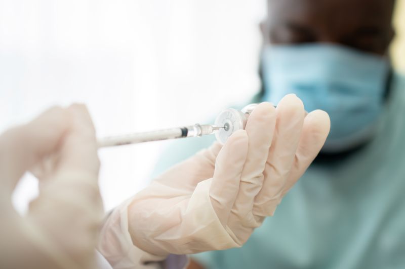
What Are Exosomes?
Exosomes are nanosized extracellular vesicles (30–150 nm) secreted by virtually all cell types. They function as the body's biological messaging system — carrying proteins, lipids, messenger RNA, and microRNA between cells to coordinate repair, regulate inflammation, and orchestrate tissue regeneration.
Unlike live cell therapies, exosomes carry none of the risks of immune rejection, ectopic tissue formation, or tumorigenicity. They are non-immunogenic, stable during storage, and capable of crossing biological barriers to reach target tissues rapidly.
HUVEC Exosomes
Derived from Human Umbilical Vein Endothelial Cells, specialized for vascular signaling, angiogenesis, and endothelial repair.
UCMSC Exosomes
Derived from Umbilical Cord Mesenchymal Stem Cells (Wharton's Jelly), specialized for broad tissue regeneration, immunomodulation, and anti-fibrotic/anti-apoptotic effects.

Why a 50/50 Combination?
No single exosome subtype addresses every challenge encountered in podiatric practice. The foot is a complex structure subjected to ischemia, neuropathy, infection, trauma, and surgical stress — often simultaneously.
A 50% HUVEC + 50% UCMSC exosome combination delivers complementary, synergistic biological signals in a single preparation — uniquely positioned to address this complexity.
The Biological Rationale
| Therapeutic Property | HUVEC Exosomes | UCMSC Exosomes | Combined Effect |
|---|---|---|---|
| Angiogenesis / Neovascularization | ★★★★★ Very Strong | ★★★★ Strong | Rapid, robust new vessel formation |
| Vascular Repair / Endothelial Integrity | ★★★★★ Specialized | ★★★ Moderate | Accelerated vessel wall restoration |
| Anti-Inflammatory (Endothelial) | ★★★★ Strong | ★★★★ Strong | Dual-pathway TNF-α, IL-1β, IL-6 suppression |
| Neuroprotection / Nerve Repair | ★★★ Moderate | ★★★★ Strong | Enhanced nerve conduction velocity, axon/myelin restoration |
| Skin Repair / Collagen Upregulation | ★★★★ Strong | ★★★★ Strong | Collagen type-1 synthesis, MMP-1 reduction, re-epithelialization |
| Multi-Tissue Regeneration (Skin, Bone) | ★★★ Moderate | ★★★★★ Very Strong | Osteogenesis, BMP-2, RUNX2 activation; cartilage & tendon repair |
| Immunomodulation | ★★★ Moderate | ★★★★★ Very Strong | M2 macrophage polarization, suppression of pro-inflammatory pathways |
| Anti-Fibrotic / Anti-Apoptotic Effects | ★★ Mild | ★★★★★ Very Strong | Reduced scarring, protected cell survival under ischemic stress |
| Oxidative Stress Reduction | ★★★ Moderate | ★★★★★ Very Strong | Critical in diabetic hyperglycemia-induced endothelial damage |
| Clinical Development Stage | Preclinical–Early Clinical | Advanced, multiple trials | Combines emerging vascular specificity with validated regenerative platform |
Key Biological Mechanisms
1. Angiogenesis and Vascular Repair
HUVEC exosomes carry a rich angiogenic cargo — VEGF, FGF, HGF, TGF-β, VEGFR-2 signaling activators, and angiogenic miRNAs including miR-210 and miR-21. In preclinical hindlimb ischemia models, HUVEC-derived exosomes increased capillary density, enlarged arteriole lumen diameter, and eliminated limb amputation compared to 43% amputation in the control group.
UCMSC exosomes contribute via miR-126, activating PI3K/Akt signaling and promoting endothelial tube formation. Together, the 50/50 combination establishes rapid, predictable neovascularization — addressing the critical barrier to healing in the ischemic diabetic foot.
2. Anti-Inflammatory and Immunomodulatory Effects
Both HUVEC and UCMSC exosomes suppress key pro-inflammatory cytokines (TNF-α, IL-1β, IL-6) through complementary pathways. UCMSC exosomes carry miR-146a, downregulating TLR-4/NF-κB signaling — a key driver of chronic inflammation in diabetic wounds. They promote M2 anti-inflammatory macrophage polarization while HUVEC exosomes restore normal vascular function in high-glucose environments.
3. Skin Repair and Collagen Upregulation
HUVEC exosomes significantly increase collagen type-1 synthesis in human dermal fibroblasts, reduce MMP-1, and promote cell proliferation — with statistically significant improvements at concentrations as low as 0.5–1% Exo-HUVEC. UCMSC exosomes reinforce this by promoting reepithelialization, ECM remodeling, and collagen deposition through TGF-β/Smad, PI3K/Akt, and NF-κB pathways.
4. Neuroprotection and Nerve Repair
Diabetic peripheral neuropathy (DPN) is a primary driver of diabetic foot ulcers and Charcot arthropathy. Exosome therapy has demonstrated significant improvements in motor and sensory conduction velocity, sciatic nerve fiber diameter, axon diameter, myelin sheath thickness, and plantar blood flow perfusion in preclinical meta-analyses.
5. Bone Regeneration and Osteogenesis
UCMSC exosomes deliver BMP-2, RUNX2, VEGF, FGF-2, and immunoregulatory microRNAs that activate osteogenic pathways. In osteomyelitis models, exosomes reduced inflammatory cytokines while upregulating osteogenic markers. In Charcot foot reconstruction, MSC exosomes reduced radiographic healing time significantly (6.4 vs. 9.2 weeks, p < 0.024) compared to surgery without MSC grafting.
Clinical Applications in Podiatric Practice
Diabetic foot ulcers carry a lifetime risk of 19–34% in diabetic patients and account for 85% of all lower-limb amputations. Standard care frequently fails due to impaired circulation, neuropathy, chronic inflammation, and infection.
A 2025 randomized controlled trial (110 patients) evaluated weekly topical application of Wharton's Jelly-derived MSC exosomes for 4 weeks in chronic DFUs, confirming MSC exosomes as a viable multimodal treatment. The addition of HUVEC exosomes in the 50/50 combination directly addresses the primary vascular deficit in DFU.
| Parameter | Guideline |
|---|---|
| Application volume | 0.1 mL/cm² of wound surface area |
| Typical dose range | 0.5–1.0 mL per treatment session |
| Frequency | Weekly for 4 weeks (followed by clinical reassessment) |
| Delivery method | Topical application to wound bed after debridement; or perilesional injection |
| Absorption | 5–10 minute absorption period before occlusive dressing |
| Follow-up | Every 2–4 weeks with Doppler assessment at baseline and 3 months |
Approximately one-third of CLTI patients are ineligible for revascularization procedures. HUVEC exosomes demonstrated zero amputations versus 43% in controls in hindlimb ischemia models, with higher capillary density and larger arteriole lumen diameter.
| Parameter | Guideline |
|---|---|
| Dose | 50–100 μg/mL per injection site |
| Volume | 1.0–2.0 mL per session, adjusted to treatment area |
| Delivery | Intramuscular injection into ischemic limb segments |
| Frequency | Series of 3–4 injections over 4–6 weeks |
| Monitoring | Pre/post Doppler ultrasound; ankle-brachial index (ABI) |
DPN is the most prevalent complication of diabetes and the primary driver of DFU, Charcot foot, and loss of protective sensation. The 50/50 combination targets DPN through UCMSC-mediated neural structure restoration and HUVEC-mediated endoneurial vascularity improvement.
| Parameter | Guideline |
|---|---|
| Delivery | Perineural injection under ultrasound guidance; or IV/IM for systemic neuropathy |
| Dose | 100–200 μg/mL concentration |
| Frequency | Every 2–4 weeks for 8–12 weeks |
| Assessment | Nerve conduction studies, monofilament testing, VAS pain scores pre/post |
MSC-derived exosomes carry anti-bacterial properties and accelerate wound repair in infected DFUs. Post-surgical application is valuable in high-risk patients undergoing debridement, skin grafting, flap procedures, or amputation revision.
| Parameter | Guideline |
|---|---|
| Volume | 0.1 mL/cm² topically to wound bed |
| Timing | Apply after surgical debridement; repeat weekly |
| Delivery | Topical spray, soaked dressing, or perilesional injection |
| Combined with | Standard antimicrobial therapy; do not apply to actively purulent wounds until infection controlled |
BMSC-derived exosomes reduce inflammatory cytokines in osteomyelitis models while upregulating osteogenic markers and VEGFA. UCMSC exosomes deliver BMP-2 and RUNX2 for bone defect repair and post-osteotomy reconstruction.
| Parameter | Guideline |
|---|---|
| Delivery | Direct injection into bone lesion/surgical site under imaging guidance; or incorporation into biocompatible scaffold |
| Dose | 100–200 μg/mL; 1–3 mL per site depending on defect size |
| Frequency | Single application at time of surgery; repeat at 4–6 weeks if required |
| Combined with | Antibiotic therapy for active infection; appropriate offloading |
Tendinopathies, ligament injuries, and osteoarthritis represent common podiatric indications for regenerative therapy. The 50/50 combination adds a vascular component ensuring regenerating tissue receives adequate blood supply.
| Condition | Dose | Delivery | Frequency |
|---|---|---|---|
| Plantar fasciitis | 1–2 mL (100 μg/mL) | Ultrasound-guided injection into plantar fascia | 1–2 injections, 4 weeks apart |
| Achilles tendinopathy | 1–2 mL (100 μg/mL) | Peritendinous injection under ultrasound | 1–2 injections, 4 weeks apart |
| Ankle osteoarthritis | 2–3 mL (100 μg/mL) | Intra-articular injection | 1–3 injections, monthly |
| Ligament injury | 1–2 mL (100 μg/mL) | Periligamentous injection | 1–2 injections, 4 weeks apart |
Charcot neuroarthropathy combines bone destruction with poor healing biology. MSC grafting reduced radiographic healing time from 9.2 to 6.4 weeks — a clinically meaningful 30% improvement. UCMSC exosomes provide osteoregenerative signals while HUVEC exosomes restore local angiogenesis.
| Parameter | Guideline |
|---|---|
| Delivery | Intraoperative injection into fusion sites and bone void |
| Combined with | Standard surgical fixation and offloading |
| Dose | 2–4 mL (100–200 μg/mL) per surgical site |
UCMSC Stem Cells: The Cellular Backbone
While exosomes provide cell-free regenerative signals, UCMSC (Umbilical Cord Mesenchymal Stem Cell) administration may be considered for cases requiring the full regenerative and differentiative capacity of living cells — particularly in large bone defects, severe Charcot reconstruction, or conditions where tissue scaffold formation is needed.
Product Comparison: Why This Combination Stands Apart
| Feature | HUVEC Exosomes Alone | UCMSC Exosomes Alone | 50/50 HUVEC + UCMSC Combination |
|---|---|---|---|
| Angiogenesis speed | Very fast, vascular-specific | Moderate-strong | Fastest, dual-pathway |
| Immunomodulation | Moderate | Very strong | Comprehensive |
| Nerve repair | Endothelial route only | Strong neural regeneration | Dual mechanism |
| Bone regeneration | Limited | Very strong | Complete osteogenic package |
| Skin/wound repair | Strong, collagen-specific | Strong, broad ECM | Synergistic, layered repair |
| Anti-fibrotic effects | Mild | Very strong | Full anti-fibrotic coverage |
| Anti-apoptotic effects | Mild | Very strong | Complete cell survival protection |
| Ischemia/PAD | Strongest available | Supportive | Leading combination for ischemic foot |
| DFU multi-factorial healing | Partial (vascular focus) | Partial (regeneration focus) | Complete: addresses all DFU failure pathways |
Dosage Reference Guide for Podiatric Applications
| Clinical Indication | Preparation | Volume/Concentration | Route | Frequency |
|---|---|---|---|---|
| Diabetic foot ulcer (DFU) | 50/50 HUVEC+UCMSC exosomes | 0.1 mL/cm² (50–100 μg/mL) | Topical to wound bed | Weekly × 4 weeks |
| PAD / CLTI / Ischemic foot | 50/50 HUVEC+UCMSC exosomes | 1–2 mL (100 μg/mL) | IM injection to ischemic segments | q2–3 weeks × 4 sessions |
| Diabetic peripheral neuropathy | 50/50 HUVEC+UCMSC exosomes | 1–2 mL (100–200 μg/mL) | Perineural injection / systemic | q2–4 weeks × 8–12 weeks |
| Soft tissue infection / post-surgical | 50/50 HUVEC+UCMSC exosomes | 0.1 mL/cm² | Topical/perilesional | Weekly |
| Osteomyelitis / bone infection | 50/50 HUVEC+UCMSC exosomes | 1–3 mL (100–200 μg/mL) | Direct bone injection / scaffold | At surgery ± repeat at 4–6 wk |
| Bone defect / fracture repair | 50/50 HUVEC+UCMSC exosomes | 2–4 mL (100–200 μg/mL) | Intraoperative into defect site | Single intraoperative + follow-up |
| Plantar fasciitis | 50/50 HUVEC+UCMSC exosomes | 1–2 mL (100 μg/mL) | Ultrasound-guided injection | 1–2 injections, 4 weeks apart |
| Achilles / tendon pathology | 50/50 HUVEC+UCMSC exosomes | 1–2 mL (100 μg/mL) | Peritendinous injection | 1–2 injections, 4 weeks apart |
| Ankle osteoarthritis | 50/50 HUVEC+UCMSC exosomes | 2–3 mL (100 μg/mL) | Intra-articular | Monthly × 1–3 sessions |
| Charcot reconstruction | 50/50 HUVEC+UCMSC exosomes | 2–4 mL (100–200 μg/mL) | Intraoperative at fusion sites | Single intraoperative |
Note: Dosage ranges presented here are derived from published preclinical and early clinical studies. Individual patient dosing should be guided by wound size, disease severity, and clinical response. Standardized clinical dosing protocols continue to evolve as the evidence base matures.
Safety Profile
- No immune rejection — exosomes are non-immunogenic and do not express HLA antigens
- No tumorigenic risk — acellular; cannot replicate or undergo malignant transformation
- No infusion toxicity — well-tolerated in multiple preclinical and clinical studies
- Stable storage — stored at −20°C to −80°C; must be used promptly after thawing
- Adverse events — the 2025 RCT of WJ-MSC exosomes in DFU (110 patients) reported no serious adverse events attributable to exosome therapy
Storage and Handling
- Store at −20°C to −80°C in the provided formulation buffer
- Thaw at room temperature or in a 37°C water bath immediately before use — do not refreeze
- Use within the specified window after thawing
- Do not mix with other biologics unless specifically validated
- Protect from light and particulate contamination
Summary: The Clinical Case for the 50/50 Combination
The challenges of podiatric medicine — particularly in the diabetic foot — are multifactorial: vascular insufficiency, neuropathy, impaired immunity, poor bone quality, and compromised skin repair occur simultaneously and reinforce each other. No single therapeutic agent addresses all these pathways.
The 50% HUVEC + 50% UCMSC exosome combination is the first cell-free regenerative platform designed to simultaneously target:
- Vascular failure — through HUVEC-mediated endothelial angiogenesis
- Chronic inflammation — through dual NF-κB suppression and M2 macrophage polarization
- Neuropathy — through neural structural repair and endoneurial vascular restoration
- Skin and wound failure — through collagen-1 upregulation, MMP-1 reduction, and re-epithelialization
- Bone and structural failure — through BMP-2, RUNX2, and osteogenic miRNA delivery
- Anti-fibrotic/anti-apoptotic protection — through UCMSC-mediated cell survival signaling
This is not a modification of existing therapy. This is a new paradigm.
Get in Touch